New technologies are helping primary care physicians more effectively diagnose diabetic retinopathy (DR), a frequent complication of diabetes, without requiring the patient to schedule a follow up referral appointment for an initial exam with an eye care professional.
Stepping back, what exactly happens to the eye when DR strikes? To fully understand the effects of the disease, it’s important to understand the structures of the eye and their functions.
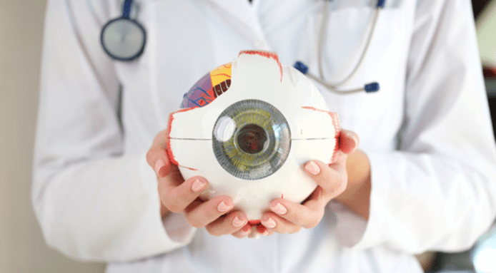
The main parts of the human eye
Many people may be familiar with the exterior of the eye, which is comprised of the pupil, lens, and cornea. However, they may be less familiar with the interior of the eye, including the fundus, or the inside, back surface of the eye, which can be negatively impacted by diabetes.
Ocular landmarks of the fundus that help us understand the effects of DR include:
Retina
The retina is the cell layer at the back of the eyeball which converts light into electric signals for the brain.
Optic disk
The optic disk is located at the back of the eye. It is a structure around the optic nerve where the sensory fibers and retinal vessels pass through the eyeball.
Macula
The macula is at the center of the retina and provides central vision, most color vision, and fine details thanks to many photo receptor cells that detect light.
Fovea
The fovea is located at the center of the retina and is responsible for central vision and fine details of objects in view.
Posterior pole
The posterior pole is important for central and peripheral vision and is located in the back of the retina. It actually includes the retina, optic nerve, the macula and vitreous humor (gel that holds the shape of the eye).
The effect of diabetic retinopathy on the eye
High blood sugar levels cause damage to parts of the eye, especially the light sensitive tissues of the retina and its related structures – this in turn can create scar tissue. Over time, the growing scar tissue pulls the retina away from the back of the eye, which interferes with vision. Impacted blood vessels can swell or leak, or even become blocked and prevent healthy blood flow. There are two types of DR:
Non-Proliferative Diabetic Retinopathy
The early stage of the disease when the retina is beginning to swell due to initial blood vessel damage.
Proliferative Diabetic Retinopathy
The more advanced stage when the retina grows new blood vessels that are prone to leaking and eventual scar tissue.
Identify retinopathy early for effective treatment
Diabetic retinopathy may not cause any symptoms initially but finding it early can help prevent vision loss and blindness.1 However, past statistics show that patients with diabetes often do not make an appointment with an eye care specialist for an eye exam for diabetes, leaving them at risk for further progression of the disease. The good news for point-of-care physicians is that it is possible to impact patient outcomes by implementing an AI diagnostic system, like LumineticsCore™ (formerly IDx-DR), that autonomously diagnoses patients for DR, making it easier for primary care physicians to identify DR at the point-of-care, possibly eliminating the need for a specialist referral.
More impactful primary care diabetic eye exams
While DR is a leading cause of blindness among working age adults, the vision loss is preventable with early intervention. Understanding the structures of the eye and their functions as well as how DR damage impacts vision can help you educate patients why having an AI powered diabetic retinopathy exams that can identify DR sooner can help you deliver the advanced care necessary to prevent vision loss in those living with diabetes.
Looking for more on this subject? Read A New Vision for Diabetic Eye Exams in Primary Care Offices.
LumineticsCore (formerly IDx-DR) is for providers to help address disparities and improve equity in care. LumineticsCore diagnostic outputs can help direct more actionable referrals to specialty care by identifying patients with diabetic retinopathy who are most likely to need timely vision saving interventions with an eye care provider. Learn more about its impacts to closing care gaps and preventing blindness.
References
1. Diabetic Retinopathy | National Eye Institute. (n.d.). Www.nei.nih.gov. Retrieved April 17, 2023, from https://www.nei.nih.gov/learn-about-eye-health/eye-conditions-and-diseases/diabetic-retinopathy#:~:text=Diabetic%20retinopathy%20may%20not%20have